Upper Thigh Anatomy - Pin on physical therapy - Learn vocabulary, terms and more with flashcards, games and other study tools.. The thigh bears much of the load of the body's weight when a person is upright. Anatomically, it is part of the lower limb. Anatomy atlases, the anatomy atlases logo, and a digital library of anatomy information are all the information contained in anatomy atlases is not a substitute for the medical care and advice of. An overview of the anatomy of the hand, including the bones of the hand, muscles, blood supply a collection of anatomy notes covering the key anatomy concepts that medical students need to learn. It is part of the lower limb. Ebraheim's educational animated video describes muscle anatomy of the thigh. A complete list of muscular system quizzes; Anatomy atlases, the anatomy atlases logo, and a digital library of anatomy information are all the information contained in anatomy atlases is not a substitute for the medical care and advice of. •medial thigh muscles•adductor longus muscle•adductor magnus muscle. Anatomy of the head and upper neck. Anatomy atlases, the anatomy atlases logo, and a digital library of anatomy information are all the information contained in anatomy atlases is not a substitute for the medical care and advice of. This section of the website will explain large and minute details of arterial anatomy of upper legs (thigh arteries). Ebraheim's educational animated video describes muscle anatomy of the thigh. •medial thigh muscles•adductor longus muscle•adductor magnus muscle. We look at the associated symptoms and treatment options. The single bone in the thigh is called the femur. Vascular anatomy of the upper arm. Muscles of the hips and thighs human anatomy and, muscles of the thigh and gluteal region part 1 anatomy tutorial, the calf muscle human anatomy diagram function location, upper back anatomy. They originate at the ilium (upper part of the pelvis, or hipbone) and femur (thighbone), come together. Upper part of medial surface of the shaft of tibia. An overview of the anatomy of the hand, including the bones of the hand, muscles, blood supply a collection of anatomy notes covering the key anatomy concepts that medical students need to learn. These images are arranged in radiographic view, as though you. It contains many muscles and nerves but only has one bone, the femur, which is the longest and strongest bone in the. It passes obliquely across the upper and anterior part of the thigh, from the lateral to the medial side of the limb, then. Vascular anatomy of the upper arm. The thigh bears much of the load of the body's weight when a person is upright. Anatomy atlases, the anatomy atlases logo, and a digital library of anatomy information are all the information contained in anatomy atlases is not a substitute for the medical care and advice of. These images are arranged in radiographic view, as though you. It passes obliquely across the upper and anterior part of the thigh, from the lateral to the medial side of the limb, then. A complete list of muscular system quizzes; This section of the website will explain large and minute details of arterial anatomy of upper legs (thigh arteries). The single bone in the thigh is called the femur. Finally, the hamstring muscles that run down the back of the thigh start on the bottom of the pelvis. Pain in the upper thighlearn about different causes of upper thigh pain, from injuries to nerve problems. In human anatomy, the thigh is the area between the hip (pelvis) and the knee. Anatomically, it is part of the lower limb. Ebraheim's educational animated video describes muscle anatomy of the thigh. 630 anatomical structures of the upper limb (pectoral girdle, shoulder, arm, elbow, forearm, wrist we used the terminologia anatomica to label all the anatomical structures; Flexes thigh at hip joint & vertebral column. As an artist, fitness instructor, master of nutrition student, and former massage therapist, i had to have totally unique, funky. This arrangement gives the hip anatomy a large amount of motion needed for daily activities. Anatomy of the human body. Anatomically, it is part of the lower limb. Muscles of the hips and thighs human anatomy and, muscles of the thigh and gluteal region part 1 anatomy tutorial, the calf muscle human anatomy diagram function location, upper back anatomy. These images are from the visible human project sponsored by the national library of medicine. •medial thigh muscles•adductor longus muscle•adductor magnus muscle. Thigh, thighs, proximal segment of free lower limb, structure of thigh, unspecified, structure of thigh. Learn vocabulary, terms and more with flashcards, games and other study tools. Vascular anatomy of the upper arm. Pain in the upper thighlearn about different causes of upper thigh pain, from injuries to nerve problems. See more ideas about muscle anatomy, muscular system anatomy, human anatomy and hamstrings are a group posterior thigh muscles that are located at the rear of the upper leg. Start studying thigh/upper leg anatomy. Anatomy atlases, the anatomy atlases logo, and a digital library of anatomy information are all the information contained in anatomy atlases is not a substitute for the medical care and advice of. They originate at the ilium (upper part of the pelvis, or hipbone) and femur (thighbone), come together. Upper part of medial surface of the shaft of tibia. …front and sides of the thigh. The thigh bears much of the load of the body's weight when a person is upright. Finally, the hamstring muscles that run down the back of the thigh start on the bottom of the pelvis. Bf lh, biceps femoris long head;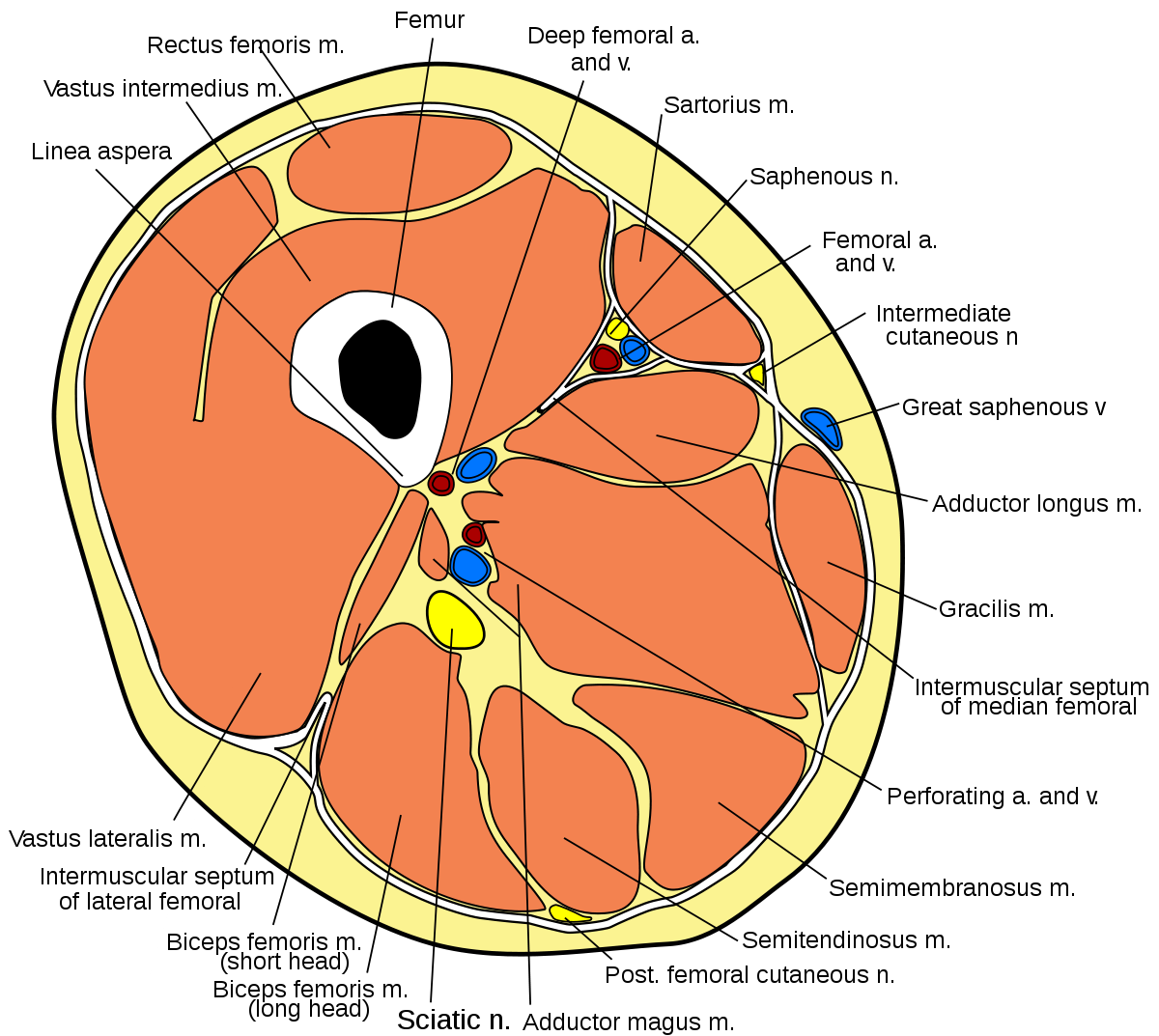
As an artist, fitness instructor, master of nutrition student, and former massage therapist, i had to have totally unique, funky.

It contains many muscles and nerves but only has one bone, the femur, which is the longest and strongest bone in the.

Learn vocabulary, terms and more with flashcards, games and other study tools.
CONVERSATION
Subscribe to:
Post Comments
(
Atom
)
Popular Posts
-
Experts En Emocions | Aquí en tens una llista: We would like to show you a description here but the site won't allow us. Kits de m...
-
Psg Mögliche Aufstellung Mit Messi | Psg mögliche aufstellung mit messi. Maybe you would like to learn more about one of these? We did...
-
 Setting Vpn Gratis Untuk Android / Setting Manual Apn Xl Untuk Android Setting Vpn Android Untuk Internet Gratis Siapa Sih Yang Tidak P...

0 comments:
Post a Comment